Home
Up

ECHONOMY
Tools for Echocardiographic Calculations

Muhamed Saric, MD, PhD
New York University


aMAZE

aMAZE Trial

Left Atrial Appendage Ligation with the LARIAT+® Suture Delivery System as Adjunctive Therapy to Pulmonary Vein Isolation for Persistent or Longstanding Persistent Atrial Fibrillation

Primary End-Points

Freedom from episodes of AF > 30 seconds at 12 months post PVI defined as:
bulletNo evidence of any episode of AF/AT/AFL > 30 seconds duration, as documented by holter monitoring or event monitor at any time following the 90-day blanking period post-index PVI through 12 months
bulletNo additional catheter ablation procedures post-index PVI for treatment of AF/AT/AFL (aside from ablation for right-sided AFL); and
bulletNo requirement for new Class I or III antiarrhythmic prescribed to treat atrial fibrillation following day 120 (90 day blanking period + 30 day window)
 

Inclusion Criteria

bulletAge ≥ 18 years and ≤ 80 years at time of screening.
bulletDocumented diagnosis of symptomatic persistent or
longstanding persistent non-valvular atrial fibrillation
bulletFailed at least one Class I or III antiarrhythmic
bulletLife expectancy ≥ 1 year;
bulletWilling and able to return to and comply with scheduled
follow-up visits and tests; and
bulletWilling and able to provide written informed consent.
 

General Exclusion Criteria

bulletPrior procedure involving opening of the pericardium or entering the pericardial space (e.g., CABG, heart transplantation, valve surgery) where adhesions are suspected
bulletPrior epicardial or endocardial AF ablation procedure;
bulletLA diameter > 6 cm as measured by CT;
bulletDocumented embolic stroke, TIA or suspected neurologic event within 3 months prior to the planned intervention;
bulletCurrently exhibits NYHA Class IV heart failure symptoms;
bulletDocumented history of right heart failure specifically when right ventricle exceeds the left ventricular size;
bulletDocumented history of myocardial infarction (MI) within 3 months prior to the planned study intervention;
bulletDocumented history of unstable angina within 3 months prior to the planned study intervention;
bulletRecent documented history of cardiogenic shock, hemodynamic instability or any medical condition in which intra-aortic balloon pump (IABP) therapy is clinically indicated;
bulletDocumented symptomatic carotid disease, defined as > 70% stenosis or > 50% stenosis with symptoms;
bulletDiagnosed active local or systemic infection, septicemia or fever of unknown origin at time of baseline screening;
bulletChronic renal insufficiency defined as eGFR < 30 mL/min/1.73m2 within 3 months prior to study treatment;
bulletEnd Stage Renal Disease (ESRD) or documented history of renal replacement / dialysis;
bulletCurrent documented history of clinically significant liver disease which predisposes the subject to significant bleeding risk (clinically defined by the treating physician);
bulletAny history of thoracic radiation with the exception of localized radiation treatment for breast cancer;
bulletCurrent documented use of long-term treatment with corticoid steroids, not including use of inhaled steroids for respiratory diseases;
bulletActive pericarditis;
bulletActive endocarditis;
bulletAny documented history or autoimmune disease associated with pericarditis;
bulletEvidence of pectus excavatum (documented and clinically defined by the treating physician);
bulletUntreated severe scoliosis (documented and clinically defined by treating physician);
bulletThrombocytopenia (platelet count < 100 x 109 /L) based on most recent pre-procedure assessment within 30 days prior to planned intervention;
bulletAnemia with hemoglobin concentration of <8 g/dL based on most recent pre-procedure assessment (within 30 days prior to planned intervention);
bulletDocumented Left Ventricular Ejection Fraction (LVEF) < 30% within 30 days prior to planned intervention;
bulletKnown acquired or inherited propensity for forming blood clots (e.g., malignancy, Factor V leiden mutation) established by prior objective testing;
bulletDocumented presence of implanted congenital defect closure devices, (e.g., ASD, PFO or VSD device);
bulletPreviously attempted occlusion of the LAA (by any surgical or percutaneous method);
bulletInability, unwillingness or contraindication to undergo TEE imaging;
bulletBody Mass Index (BMI) > 40;
bulletEvidence of active Graves disease;
bulletCurrent untreated hypothyroidism;
bulletAny contraindication to suture, endovascular device, or other minimally invasive techniques including percutaneous, transseptal, and/or sub-xiphoid access;
bulletSubject is pregnant or plans / desires to get pregnant within next 12 months;
bulletCurrent enrollment in an investigation or study of an investigational device or investigational drug that would interfere with this study and the required follow up;
bulletMental impairment or other psychiatric conditions which may not allow patient to understand the nature, significance and scope of the study;
bulletAny other criteria, medical illness or comorbidity which would make the subject unsuitable to participate in this study as determined by the clinical site Primary Investigator.
 

LAA Anatomic Exclusion Criteria

Based on screening / preprocedure imaging, subjects will also be excluded if they meet ANY of the following:

CT Criteria
Based on screening computed tomography angiography (CTA) performed within 90 days prior to study intervention as confirmed by the core lab:
bulletSuperior-posterior oriented LAA (i.e. superior C shape), that has:
bulletLAA LARIAT-approach width ≥ 40 mm; or
bulletLAA distal apex extending posterior to the ostium of the LAA.
bulletLAA positioned behind the pulmonary artery; or
bulletAll other LAA morphology: LAA LARIAT approach width > 45 mm.

TEE Criteria
Based on TEE at time of LARIAT or catheter ablation and confirmed by institution’s designated LARIAT echocardiographer:
bullet Intracardiac thrombus; or
bulletSignificant mitral valve stenosis (i.e., MV stenosis < 1.5 cm2)
 

TEE Criteria

bulletTEE Timing
bulletPeriprocedural TEE
bulletTEE at 30 days
bulletTEE at 1 year
 
bulletLeft Atrial Appendage

2D images
bullet AT LEAST the 70-90⁰, 135-150⁰ views should be obtained by single plane imaging.
bulletIdentifying the circumflex artery in short-axis and the opposing ligament of Marshall (“Coumadin ridge”) should be performed (typically from 70-90⁰)
bulletMeasurement of the maximum length of the LAA should be made (typically from 135-160⁰ view).
bulletAdditional biplane imaging may be performed.
bulletIn the setting of atrial fibrillation, long (preferably 5-10 second) captures should be performed to exclude thrombus
 

Spectral Doppler
bullet1 cm from the ostium should be recorded, optimizing the spectral Doppler velocities for accurate measurement of ejection velocities.
 

3D TEE Imaging Is MANDATORY
bulletObtain two images of the LAA; DO NOT USE MULTI-BEAT ACQUISITION (i.e. avoid splicing images); Attempt to obtain volume rates of at least 12-15 vps ; the ENTIRE LAA ORIFICE AND BODY must be imaged
bulletNarrow angle, single-beat of just the LAA
bulletFull sector, full volume (may be multi-beat) to include the entire LA and LAA


bulletLeft Atrium
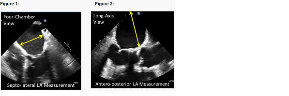
bulletFrom the 4Ch view, measure the LA septo-lateral dimension (figure 1)
bulletFrom the long-axis view, measure the LA antero-posterior dimension (figure 2).


bulletLeft Ventricle
bulletMidesophageal views
bulletLeft ventricular 4Ch, 2Ch and long-axis views should be obtained from single and/or simultaneous multiplane views
bulletTransgastric Imaging
bulletMultiple short-axis LV imaging planes (apex, mid-papillary and basal) which should include the RV
 
bulletTransseptal Imaging
bulletThe location of the orifice of the LAA and the relationship of the long axis of the LAA to the interatrial septum can be evaluated to optimize the location of the transseptal puncture. In general, a posterior-inferior fossa ovale puncture site is ideal.
bullet2D imaging from a bicaval view, using the simultaneous biplane imaging mode to determine both superior-inferior (bicaval) and anterior-posterior (orthogonal AV view) may be helpful
 
bulletMitral Valve
bullet2D - The mitral valve should be imaged from 4Ch, 2Ch and Long-axis views.
bulletColor Doppler of mitral valve using appropriate sector size to image the entire jet (Note: Nyquist limit should be between 0.5-0.6 m/s).
bulletCW of the mitral regurgitant jet to obtain a complete spectral profile of the jet


Sponsor

bulletSentreHEART; 300 Saginaw Drive, Redwood City, CA 94063