Valvular Heart Disease
Muhamed Saric MD, PhD, FACC, FASE
Director, Echocardiography Lab
Assistant Professor of Medicine
UMDNJ - New Jersey School of Medicine
Basic Anatomy of Cardiac Valves
Basic Terminology of Cardiac Valve
Pathophysiology
Basic Auscultation of Cardiac Valves
This mini-review is devoted to the anatomy, physiology and pathology of the aortic, mitral and tricuspid valves. Because isolated abnormalities of the pulmonic valve are rare in adults, disorders of this valve are not reviewed.
Basic Anatomy of Cardiac Valves
Cardiac valves have evolved in such a way to provide unimpeded unidirectional flow from the atria to the ventricles and from the ventricles to the great arteries.
The valves separating the atria from the ventricles are referred to as atrioventricular (av) valves. Those separating the ventricles from the great arteries are termed semilunar valves.
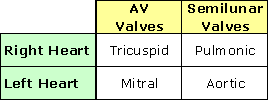
Atrioventricular valves are complex structures consisting of leaflets (or cusps) and subvalvular apparatus. The tricuspid valve has three leaflets while the mitral valve has two.
Subvalvular apparatus consists of contractile papillary muscles arising from the ventricles and nondistensible chordae tendineae, which connect the leaflets to the papillary muscles. Pulling of the chordae by papillary muscles during systole assures proper closure of the leaflets and prevents backflow of blood into the atria.
The design of semilunar valves is much simpler than that of atrioventricular valves. Each semilunar valves, whether aortic or pulmonic, consists of only three semilunar leaflets (or cusps) and no subvalvular apparatus.
With each diastole, the initial backflow of blood from the great arteries into the ventricles caused by a drop in ventricular pressure fills Sinuses of Valsalva, the pockets between the arterial wall and the semilunar leaflets. This filling shuts the semilunar valves and prevents further backflow into the ventricles.
All prosthetic valves to date (whether placed in the atrioventricular or semilunar valve positions) are imitations of the native semilunar valves.
Basic Terminology of Cardiac Valve Pathophysiology
All valvular disorders are caused by either improper opening or improper closing of the valve apparatus.
Improper opening leads to stenosis, or narrowing of the valve orifice. Stenosis impedes forward flow across the valve and imposes pressure overload on the cardiac chamber situated immediately upstream from the valve.
Stenosis of an atrioventricular valve leads to pressure overload and enlargement of the respective atrium. Stenosis of a semilunar valve results in pressure overload and hypertrophy of the respective ventricle.
Improper Valve Closure
Improper closure of either semilunar or atrioventricular valves leads to regurgitation, or valvular insufficiency. Regurgitation of any of the cardiac valves, if severe enough, may eventually lead to enlargement of the ipsilateral ventricle (e.g. aortic and mitral regurgitation) and the ipsilateral atrium (e.g. mitral regurgitation).
Severe valvular disease of the left heart, whether stenotic or regurgitant in origin, may lead to hemodynamic overload of the right heart and thus cause secondary valvular disease of the right heart. A typical example would be tricuspid regurgitation due to elevated pulmonary pressures.
On the contrary, isolated valvular disease of the right heart has no direct impact on the valvular abnormalities in the left heart.
Basic Auscultation of Cardiac Valves
Opening of normal cardiac valves creates no audible sounds. It is the closure of the valves that creates the two basic heart sounds S1 and S2.
S1 (or first heart sound) is produced by the closure of atrioventricular valves while S2 (or second heart sound) is produced by the closure of the semilunar valves.
Systole represents the interval between S1 and S2 of the same cardiac cycle. Diastole occurs between S2 of one cardiac cycle and S1 of the next.
Murmurs are abnormal sounds heard following S1 or S2. Systolic murmurs are produced by either a regurgitant atrioventricular valve or a stenotic semilunar valve. Diastolic murmurs, on the other hand, are created by either a stenotic atrioventricular valve or a regurgitant semilunar valve.
All right-sided murmurs are enhanced by inspiration.
Principles of Treatment
Antibiotic Prophylaxis
All forms of valvular heart disease predispose the patient to infective endocarditis. Therefore, all patients with significant valvular disease should receive antibiotic prophylaxis prior to medical or surgical procedures that might result in bacteremia (such as dental cleaning, intraperitoneal abdominal surgery, etc).
All forms of valvular disease are mechanical in origin and require a mechanical solution through surgical intervention. Medical management through the use of various drugs ultimately does not alter patients’ survival and is used mainly to palliate symptoms or in case of aortic regurgitation to delay the timing of surgery.
In general, surgical intervention is performed only in severe forms of valvular disease because
surgery itself leads to significant morbidity and mortality. Surgical
intervention includes:
¾ Balloon valvuloplasty for stenotic lesions (e.g. severe mitral stenosis in a young woman who is about to become pregnant)
¾ Valve repair for regurgitant lesions (e.g. mitral valve repair for severe mitral valve regurgitation)
¾ Valve replacement for either stenotic or regurgitant lesions with either tissue or mechanical valve prostheses.
There is a minimal pressure gradient across normal cardiac valves. A stenotic lesion leads to an increase in the transvalvular pressure gradient that is proportional to the decrease in valvular orifice size.
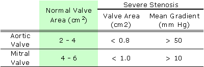
Therefore, severity of stenosis can be judged by either pressure gradient or
orifice size determined by either echocardiography or cardiac catheterization.
Patients with a stenotic valvular lesion usually do not develop symptoms unless
at least 50% of valve orifice area is lost.
Aortic Stenosis
Etiology
Most forms of aortic stenosis are cause by calcific degeneration of either congenitally bicuspid or congenitally tricuspid aortic valve.
Bicuspid aortic valve is the most common form of congenital heart disease in adults and occurs in 1-2% of all life births. Unlike the normal trileaflet aortic valve, a bicuspid valve has only two semilunar cusps. This abnormal architecture of the bicuspid valve often leads to aortic stenosis, which may become severe by age 50.
Calcific degeneration of the initially normal trileaflet aortic valve usually leads to severe stenosis decades later that the bicuspid valve and is the most common form of aortic stenosis in the elderly.
Mild and moderate forms of aortic stenosis usually produce no symptoms. The classic symptom triad of severe aortic stenosis includes angina, congestive heart failure and syncope (which may result in sudden death).
¾ Valve replacement should be offered to all patients with severe symptomatic aortic stenosis.
¾ Valvuloplasty should be reserved only for patients who cannot undergo cardiac surgery (e.g., an elderly patient with severe aortic stenosis who needs hip surgery for fractured femur).
Saric, M. and Kronzon, I. (2000) Aortic stenosis in the elderly. The American Journal of Geriatric Cardiology 9:321-329;345.
Mitral Stenosis
Etiology
¾
rheumatic heart
disease is the most common cause of mitral stenosis worldwide. Because
rheumatic heart disease has been for the most part eradicated from the
developed countries, mitral stenosis is rarely encountered in native-born US
citizens. The patient with rheumatic mitral stenosis one is most likely to
encounter in the US would be a young or middle-aged woman who immigrated from a
country where rheumatic valve disease is still prevalent.
¾ Uncommon causes of mitral stenosis include:
o Mitral annular calcifications (e.g. accumulation of calcium phosphate along the atrioventricular groove in patients with end-stage renal disease)
o Congenital abnormalities (e.g. a supravalvular membrane stretched across the atrial side of the mitral valve)
¾ Exercise Intolerance & Heart Failure: Mitral stenosis impedes the diastolic filling of the left ventricle across the mitral valve. Since tachycardia shortens the diastole, patients develop signs of heart failure (such as shortness of breath) especially during exercise.
¾
Atrial
fibrillation: Through pressure overload, mitral stenosis causes left
atrial enlargement, which then predisposes the patient to atrial fibrillation,
a rapid irregular heart beat. Atrial fibrillation is dangerous for two reasons:
o Tachycardia: The rapid heart rate induced by atrial fibrillation further worsens the symptoms of heart failure.
o
Thromboembolism:
Atrial fibrillation in conjunction with left atrial enlargement is a risk
factor for clot formation in e.g. left atrial appendage. Pieces of the clot can
embolize into the systemic circulation causing a stroke, for example.
¾ Auscultatory findings: Loud S1, opening snap following S2 and diastolic rumble following opening snap.
Medical therapy:
¾ Antibiotic prophylaxis prior to bacteremia-inducing medical and surgical procedures
¾ Heart rate-controlling agents (such as digoxin or beta-blockers)
¾ Long-term anticoagulation to prevent thromboembolism in patients with atrial fibrillation
Surgical Therapy:
¾ Valvuloplasty should be offered to all patients with symptomatic mitral stenosis whose valve anatomy is amenable to balloon valvuloplasty. Unfortunately, the effects of valvuloplasty are not permanent and mitral stenosis worsens again overtime.
¾ Valve replacement should be offered to all patients with severe symptomatic mitral stenosis who are not candidates for balloon valvuloplasty.
When compared to stenotic lesions, the severity of valvular regurgitations is much more difficult to measure objectively. The most common echocardiographic and cardiac catheterization methods rely on an estimate of the regurgitant blood volume (either visually or quantitatively).
Only recently, methods have been developed to measure the actual regurgitant orifice size. The larger the regurgitant orifice size, the more severe the regurgitation is.
Aortic Regurgitation
Etiology
¾ Leaflet abnormalities
o Congenitally bicuspid valve (Note that bicuspid valve can lead to either aortic stenosis, aortic regurgitation or combination thereof).
o Infective endocarditis
o Rheumatic heart disease (Uncommon in the Unites States. Mitral stenosis + aortic regurgitation is the classic combination seen in rheumatic heart disease).
¾ Aortic root dilatation
o Genetic & autoimmune disorders (e.g. Marfan syndrome, ankylozing spondylitis, psorisis)
o Annuloaortic ectasia (due to hypertension)
o Aortic dissection (a blood flow-driven cleavage through the aortic media in the longitudinal direction)
Acute severe aortic regurgitation is usually caused by either infective endocarditis of aortic dissection. It presents with fulminant heart failure and is often fatal if left untreated. Because of its short duration, the left ventricle is usually not dilated. The murmur may not be prominent.
Chronic severe Aortic regurgitation develops over decades and is often characterized by a massively enlarged yet dysfunctional left ventricle. Patients often complain of shortness of breath and angina (even in the absence of significant coronary artery disease).
It is characterized by a diastolic murmur best heard along the left sternal border.
Acute severe aortic regurgitation requires prompt valve surgery.
For chronic forms of the disease, one may utilize:
¾ Medical therapy: It is used primarily in patients with asymptomatic aortic regurgitation who still have normal left ventricular function. Vasodilators (such as nifedipine) delay the need for valve surgery.
¾
Valve
replacement with tissue or mechanical prosthesis should be offered to
all patients with severe aortic regurgitation who are either symptomatic or
have left ventricular systolic dysfunction.
Mitral Regurgitation
Etiology
¾ Mitral Annular Dilatation
o Left ventricular dilatation of any cause such as dilated cardiomyopathy, or severe aortic regurgitation.
¾ Excessive Leaflet mobility
o Mitral Valve Prolapse: This is the most common form of mitral valve disease in the United States requiring valve surgery. In this disorder mitral valve leaflets and chordae undergo myxomatous degeneration and lengthening which eventually leads to mitral valve regurgitations.
¾ Restrictive leaflet mobility
o Rheumatic heart disease
o Papillary muscle displacement due to ischemic heart disease (e.g. ischemic mitral regurgitation following infarction of the inferior left ventricular wall)
Acute severe mitral regurgitation presents with fulminant heart failure and is often fatal. Because of its short duration, left atrium and left ventricles are usually not dilated. The murmur may not be prominent.
Chronic severe mitral regurgitation develops over decades and is often characterized by a massively enlarged yet dysfunctional left atrium and left ventricle (the so-called cor bovinum, or bovine-sized heart).
It is characterized by a holosystolic murmur best heard at the cardiac apex.
Acute severe mitral regurgitation requires prompt valve surgery.
For chronic forms of the disease, one may utilize:
¾ Medical therapy: Palliates symptoms of heart failure but does not prolong survival.
¾ Valve repair (most commonly performed for mitral valve prolapse)
¾
Valve
replacement with tissue or mechanical prosthesis should be offered to
all patients with severe mitral regurgitation who are either symptomatic or
have left ventricular systolic dysfunction.
Tricuspid Regurgitation
Etiology
¾ Secondary tricuspid regurgitations: This is the most common form of tricuspid valve disease and develops in response to chronically elevated pulmonary artery pressures in e.g.:
o Left-heart failurE of any cause
o
Chronic
obstructive pulmonary disease (COPD)
of smokers
¾ Primary tricuspid regurgitation
o Infective endocarditis (often associated with intravenous drug abuse or medical devices such as catheters and pacemaker wires).
o Carcinoid tumors of the gut secrete humoral factors that case stiffening of tricuspid leaflets which, in turn, leads to both tricuspid stenosis and tricuspid regurgitation
o Congenital abnormalities (such as Ebstein’s anomaly in which the tricuspid valve is displaced toward the cardiac apex).
Tricuspid regurgitation, if severe enough, ultimately leads to right-heart failure manifested by lower extremity edema and ascites.
Because most causes of tricuspid regurgitation are secondary to left-heart or lung disease, the therapy is directed toward the underlying cause.
Tricuspid valve replacement has a high complication rate (including prosthetic thrombosis) and is rarely done. Tricuspid valve annuloplasty is the most common form of tricuspid valve repair.